No. 019
Human Skull Facts: Why Skulls Have No Nose, and Other Things to Know
A skull has no nose because most of the nose was never bone. The part that sticks out from your face is cartilage, skin and other soft tissue, and none of that lasts once a body becomes a skeleton. What stays is the bony bridge at the top of the nose and the nasal cavity behind it, which is why every skull, real or plastic, has that dark, upside-down heart shape in the middle of its face. The outer ear goes the same way, since it is mostly skin and elastic cartilage. What the skull does keep is plenty: 22 bones, usually a set of teeth, and the case that once held a brain.
If you have just stood in front of a skull in a museum case, sketched one from a textbook, or set a grinning prop on the porch for October, the questions tend to come in the same order. Where did the nose go? How many pieces is this thing made of? Why are the teeth still there? The answers below take the parts people ask about most, one feature at a time.
Where the nose and ears went
Start with the face, since that is where the skull looks most unlike the person it belonged to. According to Visible Body, the two small nasal bones form only the bridge of the nose, the hard ridge you can feel between your eyes. Below that, nasal cartilage forms most of the shape that projects outward, including the tip. Cartilage is tough and flexible in life, but it is not bone, so it does not survive on a dry skull.
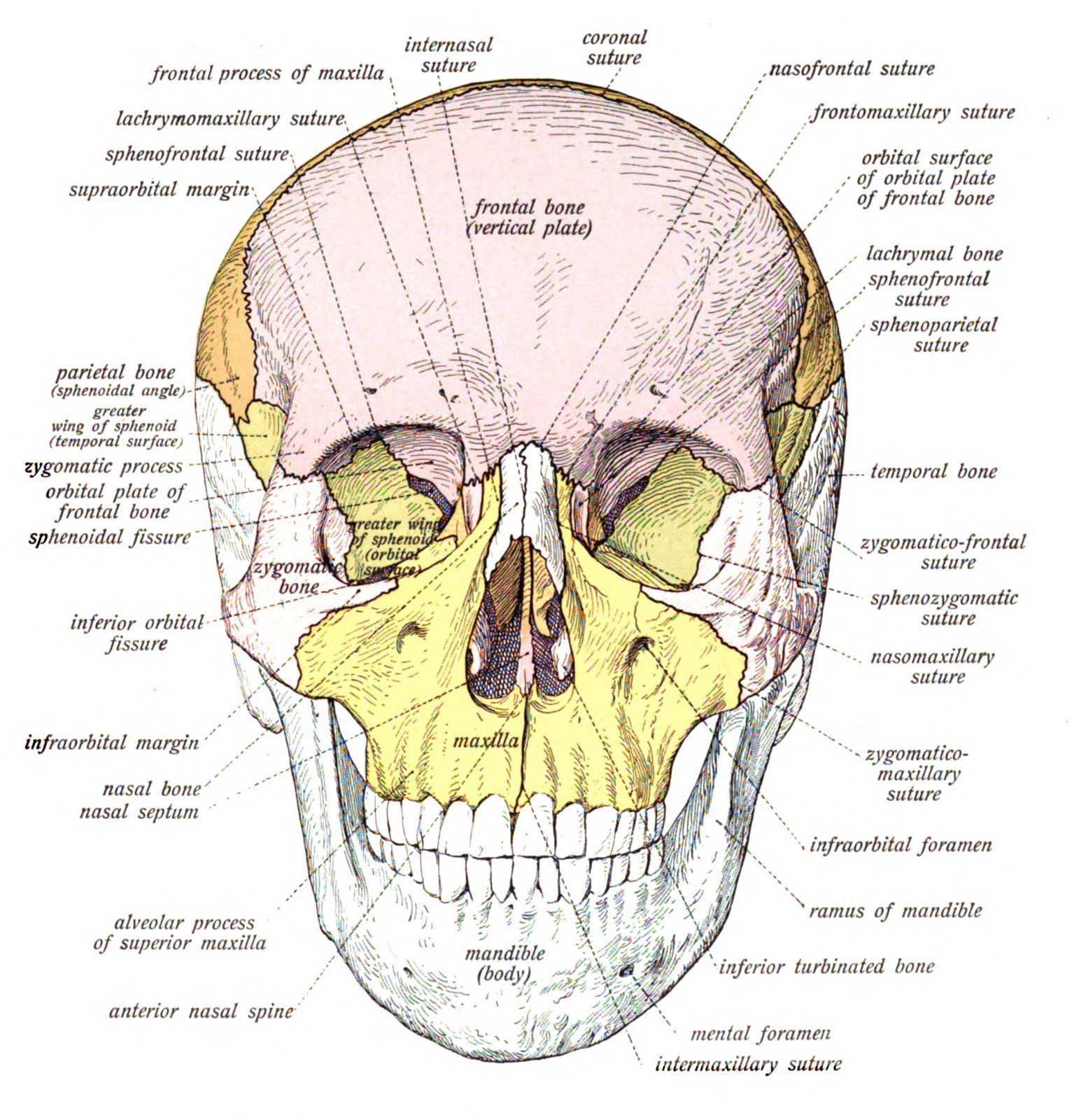
The skull still holds a lot of nose, just not the part anyone draws on a face. Cleveland Clinic lists the bony nasal cavity, the nasal bones, the vomer (a thin bone that forms part of the wall dividing the nasal cavity) and parts of the ethmoid and maxilla. That is the opening you see on a skull, the entrance to the airway rather than the nose itself.
Ears follow the same rule. The visible outer ear, called the auricle, is mainly skin and elastic cartilage, so it is missing from a skeleton too. The bony ear canal stays, opening on each side of the skull through the temporal bones. Deeper inside those temporal bones sit the smallest bones in the body, the malleus, incus and stapes of the middle ear. So, a skull without ears still has its hearing hardware tucked away; you just cannot see it from the outside.
How many bones make up a skull
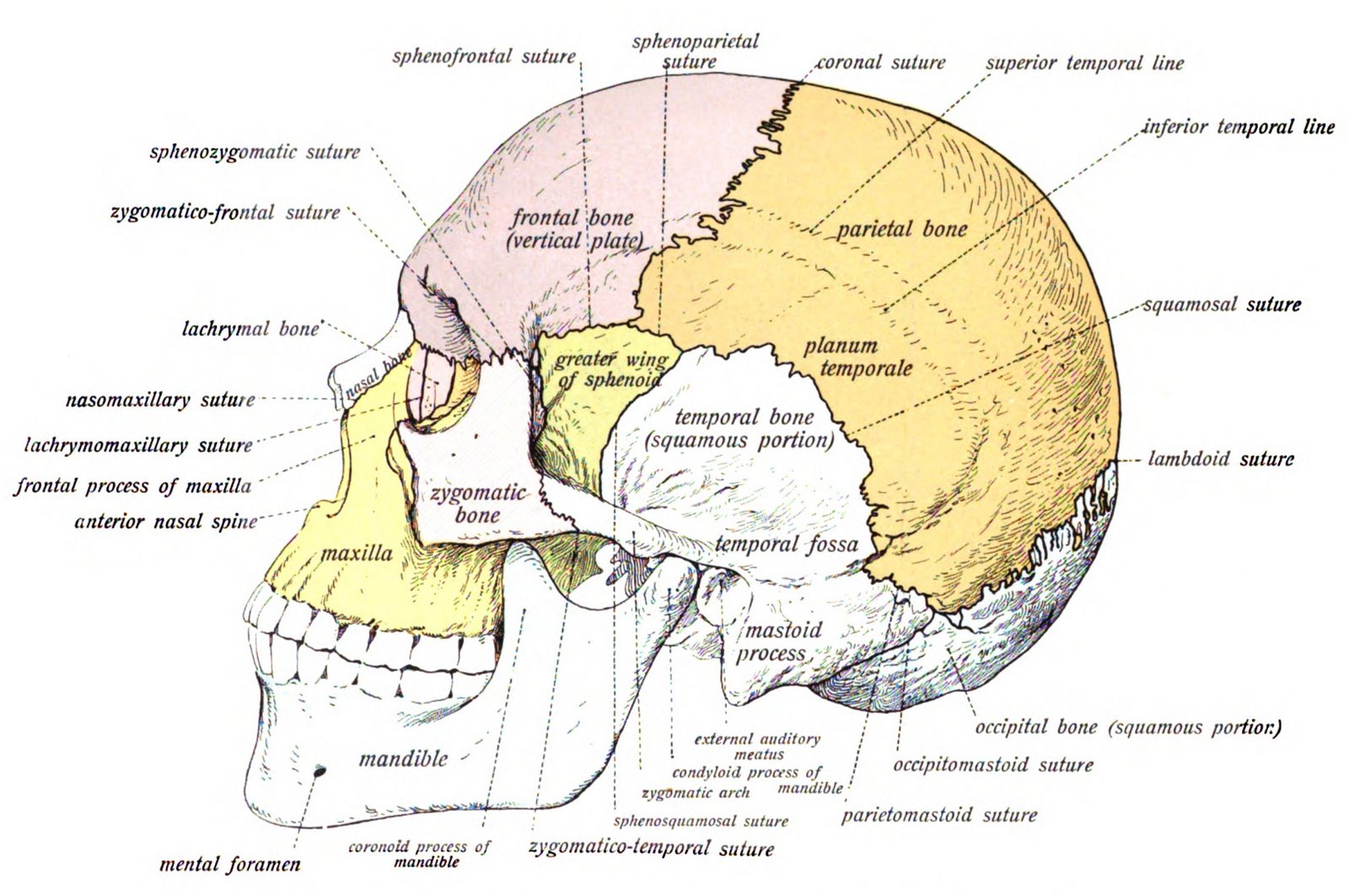
A skull looks like a single bowl of bone, but it is a puzzle of many pieces fitted tightly together. The adult human skull has 22 bones, according to Cleveland Clinic: eight cranial bones that surround the brain and 14 facial bones that build the face.
Tap or hover a row to highlight it.
| Group | Count | Bones |
|---|---|---|
| GroupCranial bones | Count8 | BonesFrontal, two parietals, two temporals, occipital, sphenoid, ethmoid |
| GroupFacial bones | Count14 | BonesTwo maxillae, two zygomatics, mandible, two nasals, two palatines, two inferior nasal conchae, two lacrimals, vomer |
| GroupTotal skull bones | Count22 | BonesCranial bones plus facial bones |
The cranial bones are the ones you would point to on a drawing of a head. The frontal bone forms the forehead. The two parietal bones make up most of the top and sides. The two temporal bones sit at the sides near the ears, and the occipital bone curves around the back and base. The sphenoid and ethmoid are harder to see from outside, because they sit deep in the middle of the skull behind the eyes and nose.
The facial bones include the two maxillae of the upper jaw, the two zygomatic bones that form the cheekbones, and the mandible, the lower jaw. Smaller pieces fill in around the nose and eye sockets: the nasal bones, the palatine bones at the back of the roof of the mouth, the inferior nasal conchae inside the nasal cavity, the tiny lacrimal bones in the inner corner of each eye socket, and the vomer.
Two things are left out of that count. The teeth are attached to the maxilla and mandible, but they are not counted among the 22 skull bones. The six middle-ear bones are usually counted separately as well. If you want the full tally for the rest of the body, the guide to how many bones are in the human body picks up where the skull leaves off.
The one bone that moves
Here is a skull fact worth keeping for the next trivia night: the lower jaw is the only part of the skull that moves. Cleveland Clinic describes the mandible as the skull's one movable bone, hinged at the temporomandibular joints just in front of the ears. Every other main bone of the skull is locked to its neighbors by sutures or other fixed joints.
That is why anatomy models and many Halloween skeletons have a jaw on a spring or a wire. The cranium is one solid unit, and the mandible is the separate piece that opens and closes for chewing and speaking. A skull that seems to be laughing at you is simply showing off the only joint it has.
What the skull protects
The skull's main job is protecting the brain. Cleveland Clinic adds that it also supports and shields the sensory organs, such as the eyes and ears, which is why the eye sockets are deep bony cups and the inner ear is buried in some of the densest bone in the head.
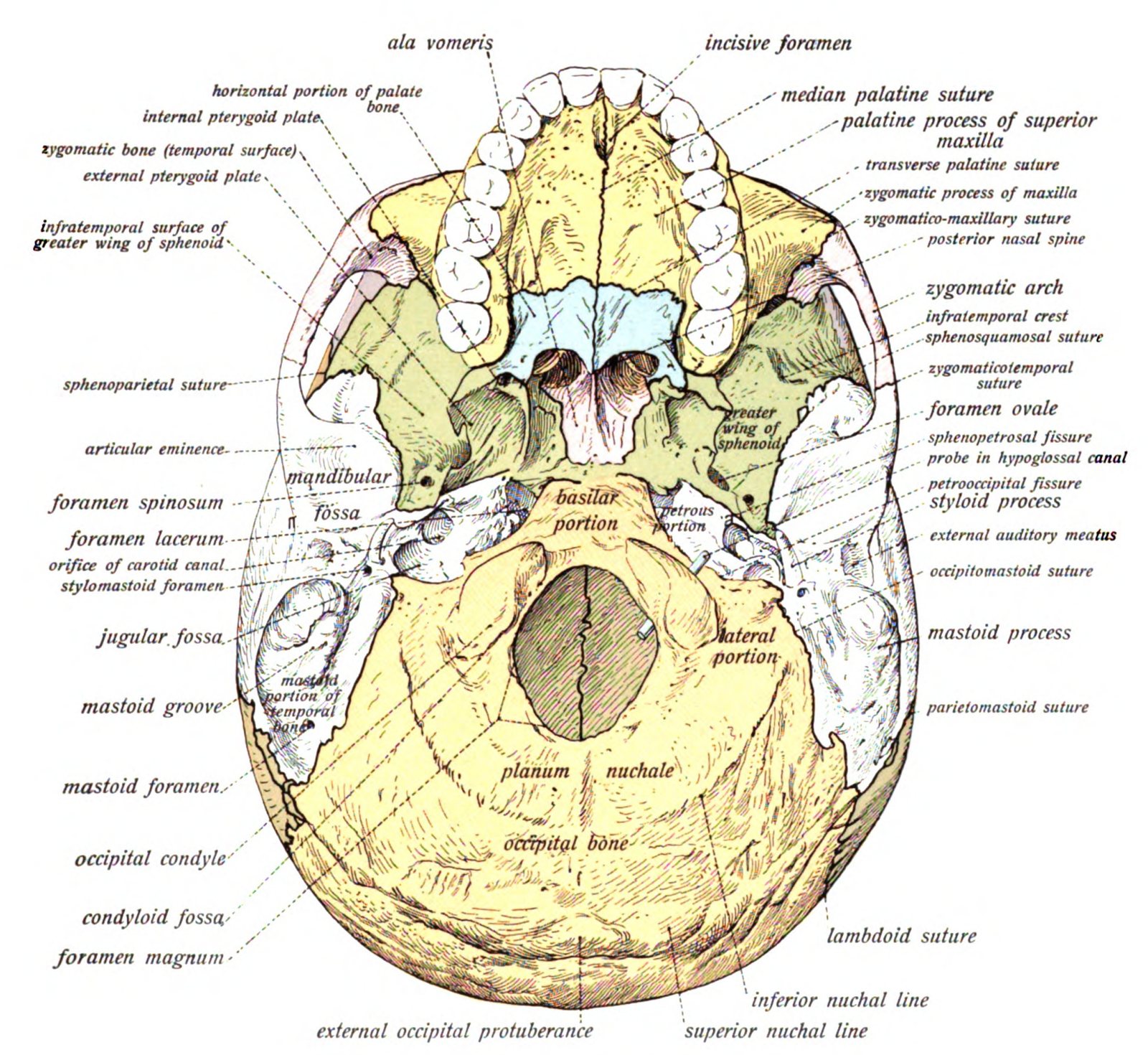
Turn a skull over and you will see the biggest hole in it, at the base. This is the foramen magnum, Latin for "great opening." Visible Body describes it as the largest opening in the skull, the place where the spinal cord and the vertebral arteries pass through. Smaller openings, called foramina, are scattered across the base for nerves and blood vessels, so a skull is less a sealed box than a well-guarded case with carefully placed doorways.
Why the teeth stay in
Teeth are not bones of the skull, so it surprises many people that they are still in place on so many skulls. The reason is in how they are anchored. Each tooth root sits in a socket in the maxilla or mandible. A network of connective-tissue fibers called the periodontal ligament attaches the cementum, the hard coating on the root, to the bone lining the socket, according to the NCBI Bookshelf.
Teeth can go missing for two very different reasons. During life, teeth are lost to decay, periodontal disease, injury or extraction, a point the American Dental Association makes about gum disease in particular. After death, the periodontal ligament breaks down, and teeth can loosen in their sockets. A study of evidence from teeth and the bone of skulls lists other factors that affect whether they stay: exposure to the elements, handling, excavation, scavenging animals and the shape of the roots.
The empty socket tells you something too. When a tooth was lost during life, the bone around the socket may remodel or heal over. When a tooth fell out after death, the socket usually keeps a sharp, unhealed edge. That is one reason a skull with gaps in its smile can still hold a little of its owner's story.
A baby's skull: soft spots and seams
A newborn's skull is built to change shape. Its bones have not yet joined, and the spaces between them are covered by membranes. These are the fontanelles, the soft spots on a baby's head. Cleveland Clinic explains that they let the head mold slightly during birth and give the brain room to grow afterward.
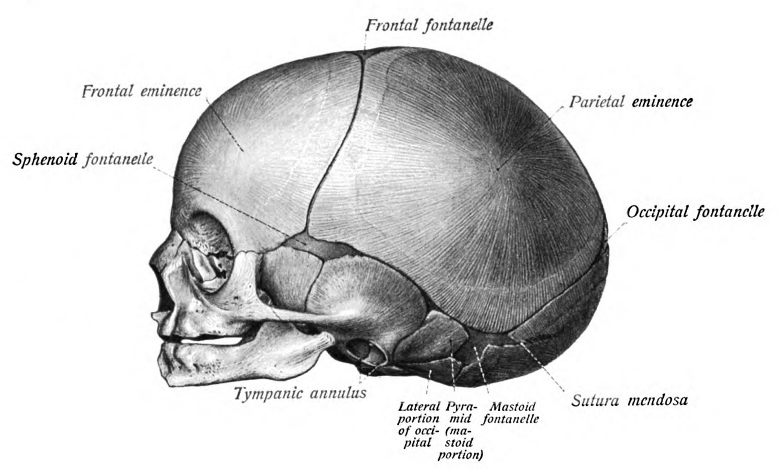
A baby has two well-known fontanelles. The posterior fontanelle, near the back of the head, usually closes around six to eight weeks. The anterior fontanelle, the larger soft spot near the front, usually closes around 12 to 24 months. Between the bones run sutures, fibrous joints with names that are fun to say out loud: coronal, sagittal, lambdoid, metopic and squamous. They stay flexible in infancy and childhood, then gradually stiffen and may fuse, each on its own timetable. The NCBI Bookshelf notes that several major sutures keep closing into early adulthood, and the timing varies from suture to suture and from person to person.
How a skull grows and closes
Tap or hover a step to follow the order.
Birth
Fontanelles and flexible sutures allow molding and brain growth
About 6 to 8 weeks
Posterior fontanelle usually closes
About 3 to 9 months
Metopic suture commonly fuses
About 12 to 24 months
Anterior fontanelle usually closes
Early adulthood
Many major sutures continue toward fusion at different times
These ages are typical ranges, not a checklist for any one child. If you have a question about a particular baby's soft spot or head shape, the pediatrician is the person to ask.
The weakest spot, and the toughest areas
The skull's best-known weak point has its own name: the pterion. It sits on each side of the head, roughly at the temple, where four bones meet in an H-shaped junction: the frontal, the parietal, the squamous part of the temporal, and the greater wing of the sphenoid. Oregon State University's anatomy text explains why it gets the label. The bone is relatively thin there, and the anterior branch of the middle meningeal artery runs just underneath it. Anatomists have studied its exact position and thickness closely, as in a paper on the surface anatomy of the pterion, because it matters so much in medicine.
The strong side of the story has no single winner. Anatomy texts point to a few especially sturdy regions instead. The frontal bone is thickened above the eye sockets at the brow ridges. At the base of the skull, the petrous part of the temporal bone forms a dense ridge that houses the structures of the middle and inner ear. Those are the areas built heaviest, and they show how the skull puts its thickest bone around the parts that need it most.
None of this is a guide to head injuries, which belong with a doctor. It is simply a map of how the skull is put together.
If you have read this far, these five should be easy.
Human skull quiz
Answer every question to check.
Questions people ask about the human skull
The same few questions come up again and again, in English and in Spanish, so the short answers follow.
Why do skulls have no nose?
Skulls have no nose because the part of the nose that projects from the face is mostly cartilage, skin and other soft tissue, which does not last on a skeleton. The skull keeps only the nasal bones at the bridge and the bony nasal cavity behind them. That is why a skull shows an opening where the nose used to be.
Why do skeletons have no ears?
Skeletons have no ears because the outer ear, the auricle, is made mostly of skin and elastic cartilage rather than bone. The bony ear canal stays, and the three tiny middle-ear bones remain hidden inside the temporal bones on each side of the skull.
What is the weakest part of the skull?
The pterion is traditionally called the weakest part of the skull. It is the H-shaped meeting point of four bones on each side of the head, where the bone is relatively thin and the middle meningeal artery lies just beneath, according to Oregon State University.
What is an interesting fact about the human skull?
One favorite is that the mandible, the lower jaw, is the only movable bone in the skull. Another is that a baby's skull starts out with soft spots that close over the first two years, while some of its seams keep closing into early adulthood.
What organ does your skull protect?
Your skull mainly protects your brain. It also supports and shields sensory organs such as the eyes and ears, and its largest opening, the foramen magnum, lets the spinal cord connect to the brain.
Where to go next
Anatomy is only half of why people love skulls. For what the skull has meant to artists, churches and sailors over the centuries, read about skull symbolism, and for the old idea of the skull as a nudge to live well, see what memento mori means. If you would rather put pencil to paper, the guide to how to draw a skull turns those cheekbones and eye sockets into lines. And if the skull that started all this was a giant plastic one in a neighbor's yard, meet the 12-foot skeleton.
For the people who know the inside of a body best and keep the lights on after midnight, our Night Shift Nurse skull tee puts an engraved skull in a nurse's cap and stethoscope under a crescent moon, with CALM AFTER MIDNIGHT across a marigold ribbon.